Aiken, SC
info@equus-soma.com
Equus-Soma
Equine Osteology & Anatomy Learning Center
Waldoboro, ME
207-542-6132
Front Hooves
Apollo's Front Limb Coffin Bones
As mentioned in Part One, our primary plan when recovering Apollo was to focus on getting his axial skeleton and not necessarily his legs. We did an on-site, precursory examination of the long bones (of the legs) but since we did not see any obvious pathologies we decided not to keep them. A little later, Eva mentioned that Apollo had had issues with his front hooves so we returned to the dig and retrieved the distal bones of both front feet which included the carpal (knee), cannon, splint, sesamoids, long pastern (P1), short pastern (P2), navicular and coffin (P3) bones.The most note-worthy pathology was found in both the left & right coffin bones which correlated with Eva's recollection that Apollo was plagued with numerous abscesses and a keratoma.
- "Summer of 2018 also brought on the abscesses and the keratoma ... switched to (a new farrier) who was amazing, and glue on shoes. His front feet just stopped growing no matter what hoof supplement he was on."
~Eva McGuire
"A keratoma is a benign tumor of the inner layer of keratin-producing epidermal hoof wall cells that forms inside a horse's foot. As the tumor slowly grows, it expands and separates the hoof wall laminae, causing pain and lameness."
"When the tumor grows down to the sole, separating the white line, infection may gain access (leading to abscess formation)."
From the web search: www.VCAhospitals.com

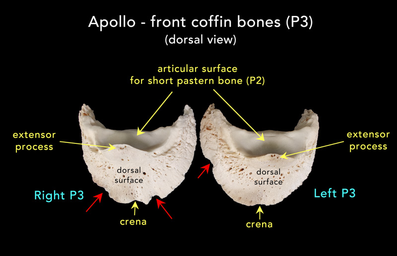
Above Photo: Right and Left coffin bones. The red arrows indicate areas of bone demineralization due to abscessing and a keratoma (deeper crescent-shape indentation on right P3). The "crena" is a normal, shallow indentation at the toe of most coffin bones.
The four images below illustrate the medial and lateral views of both right and left coffin bones. Of particular note are the areas within the red square and circles where we see irregular pores and enlarged cavities in the region distal to and including the palmar processes.
The proximal (upper) border of each palmar process attaches to the lateral cartilage. In the "normal" situation, the surfaces of these processes are relatively smooth (not so in Apollo's case). See Apollo ref #29.**Click on any image to ENLARGE**
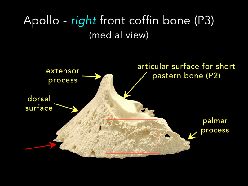
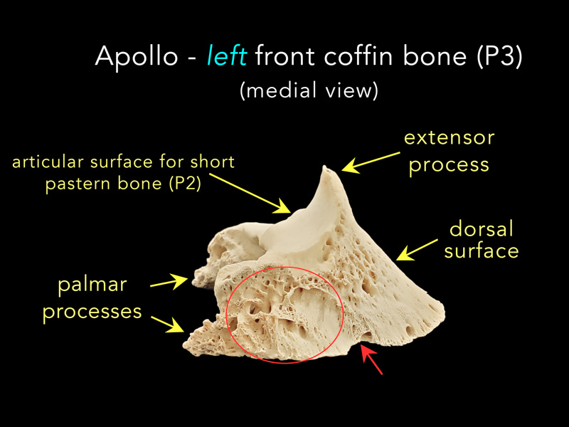
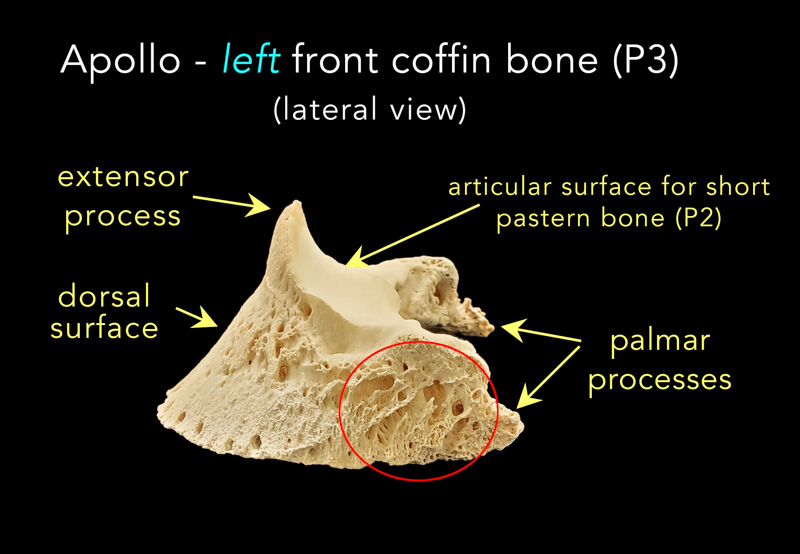
PHOTO CREDITS: The majority of images used on this website are property of Equus-Soma (Pamela Blades Eckelbarger). Images of me taken at Presentations are provided courtesy of Helen Peppe and other attending participants (thank you!!). Images on the About page of myself competing with Irish are courtesy of Flatlandsfoto. Images of skeletons in the banners are from Muybridge 1881.

November to July
1165 Shaws Fork Rd.
Aiken, SC 29805Equus-Soma
Equine Osteology & Anatomy Learning Center
Pamela Blades Eckelbarger M.S. Zoology
eqsoma71@gmail.com
(207) 542-61322025 ©ALL RIGHTS RESERVED
July through October
190 Horscents Ln.
Waldoboro, ME 04572