Aiken, SC
info@equus-soma.com
Equus-Soma
Equine Osteology & Anatomy Learning Center
Waldoboro, ME
207-542-6132
Apollo's Story - What Lies Beneath - Part One
Background
After making the heart-wrenching decision to humanely euthanize her 8 yr old OTTB "Apollo" in October 2018, his owner Eva McGuire asked if I would recover his bones to see if they might provide some answers to the dramatic change in his behavior which had become difficult and sometimes dangerous in the last year of his life. Apollo's body was buried (above ground) at Compassionate Composting in Auburn Maine and we exhumed his bones in October 2019.Our very preliminary examination of Apollo's axial skeleton (spine) revealed some interesting pathologies about which Eva posted on her personal Facebook page (10/23/19). Within days her post went "viral" with 10,000 'likes' and 13,000 shares. There have been hundreds of comments and questions but also false assumptions based on the few photos of the bones she posted. Eva then started a page dedicated to his story "Apollo and the story of his bones" where she has shared detailed accounts of Apollo's short time with her and everything she did to find answers to help him.
With the hope of having a little more control over the multitude of responses and wayward comments but mainly wanting to limit membership to those seriously interested in this study, we also created a private Facebook Group "Apollo - The search for answers in his skeleton".
- "One of the very important reasons for me to do this is that I wanted people to see Apollo first as a breathing, living, playful beautiful being."
"I want people to realize his journey, and the many, many ups and downs ... and what led to the end."~ Eva McGuire
The Dig
When hearing of Apollo's behavioral changes our first suspicion was that he may have had ECVM, a congenital malformation that involves the last two cervical (neck) vertebrae, the first thoracic (back) vertebra, first pair of sternal ribs and/or the associated soft tissues. (Please refer to the ECVM page for details and photos.)
As our focus was on looking at Apollo's cervical vertebrae, we uncovered his skull & neck first. Surprisingly, Apollo did not have ECVM so we continued to collect the rest of his axial skeleton. We did an on-site, precursory examination of the long bones (of the legs) but not seeing any obvious pathologies we decided not to keep them (space has become very limited at the Learning Center!). A little later, Eva mentioned that Apollo had had issues with his front hooves so we returned to the dig and retrieved the distal bones of both front feet.
Our intention here is to document the examination of Apollo's bones. We will illustrate what we suspect to be anomalies and clinical pathologies based on similar irregularities previously described in peer-reviewed publications.
** CLICK ON ANY IMAGE TO SEE A LARGER VIEW**
Equine Anatomy 101
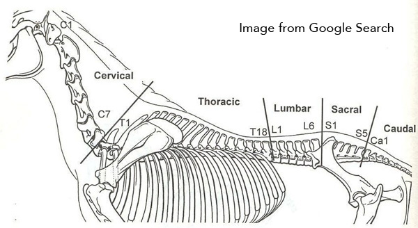
Before launching into what is becoming a rather complex study of Apollo's bones, it is important to have a basic understanding of how the equine axial skeleton is designed (see Apollo ref. #20).
The most common "vertebral formula" in the horse is: C7 (seven cervical or neck), T18 (eighteen thoracic or back), L6 (six lumbar), S5 (five sacral) and Ca 18-20 (caudal or tail) vertebrae.The number of cervical vertebrae (7) is constant in horses, whereas variations in the numbers of thoracolumbar (thoracic + lumbar) vertebra are not uncommon (see Apollo ref. #12). In the collections at the Learning Center for example, we have a Friesian with 19 thoracic vertebrae, several horses with 5 lumbar vertebrae including our "Founding Father" Petey, as well as, a few others with only 4 lumbar vertebrae.
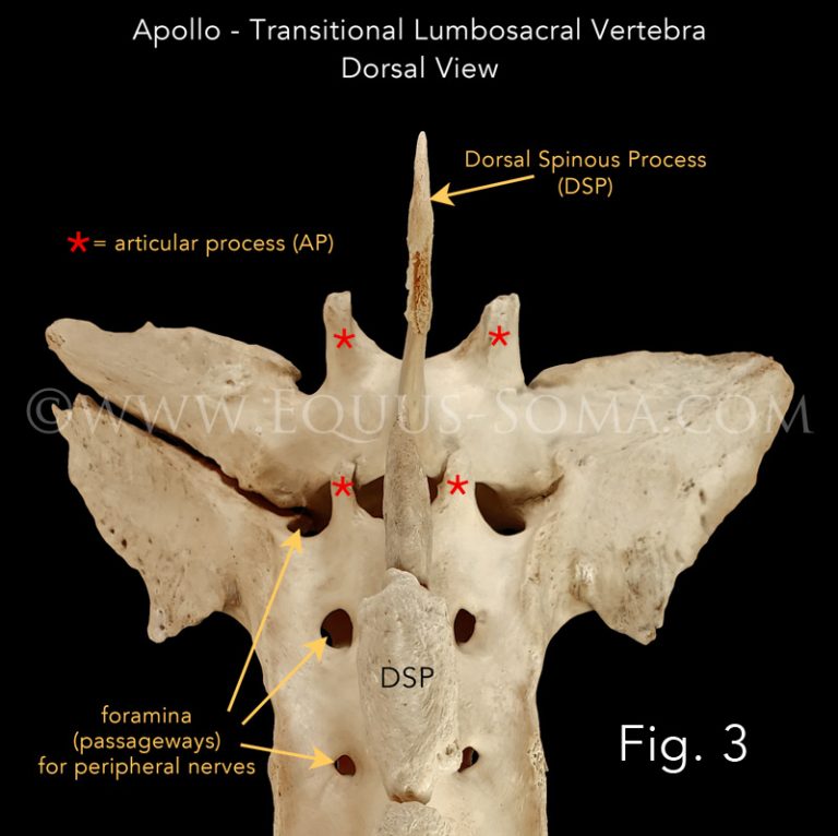
Unlike thoracic vertebrae, lumbar vertebrae do not bear ribs. They are characterized by having flat, "wing-like" transverse processes. The lumbar region of the spine (behind the saddle) is not built to carry weight. It was designed for stability and protection of the underlying viscera.The lumbar transverse processes provide attachment sites for surrounding muscles including the very important psoas major and minor muscles that run along the ventral surfaces.

Apollo's Story - Part One: The Beginning of the End?
Apollo's physical troubles likely started before he was born. Specifically, during the embryological stages of vertebral development. Apollo was born with a "transitional lumbosacral vertebra" which is unusual but not uncommon (see Apollo refs. 5, 9, 12,14, 15 & 19).
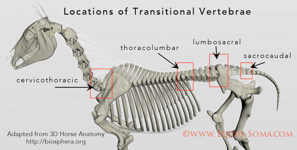
Transitional vertebrae (also found in humans & other mammals) are located between two adjacent spinal regions i.e., between the neck & thorax (cervicothoracic), thorax & lumbar (thoracolumbar), lumbar and sacrum (lumbosacral) and between the sacrum & caudal (tail) (sacrocaudal) vertebrae. Transitional vertebrae exhibit physical characteristics of both the vertebra immediately before and the one after as a result of faulty gene expression (see Apollo ref. 11).
Apollo entered the world with his 6th lumbar vertebra (L6) partially fused with the 1st sacral vertebra. This type of transitional configuration is referred to as the "sacralization of L6" (see Apollo refs. 9, 12, 14 & 19).
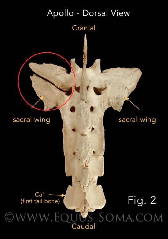
In Apollo's case, the right sacral wing is fused with the right transverse process of the last lumbar vertebra (L6) but the left sacral wing is not fused with the left transverse process of "L6" (BELOW Figs. 2 & 5 - red circle), resulting in asymmetry in not only the lumbosacral joint, but the sacroiliac joint (SI) as well.
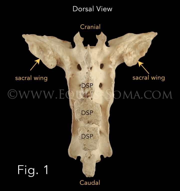
The photos below compare the morphology of a "typical" sacrum (Figs. 1 & 4) with that of Apollo's sacrum (Figs. 2, 3, 5 & 6). A "typical" sacrum has 4 or 5 vertebrae that are fused at the vertebral bodies.. The first sacral vertebra is characterized by expansive, relatively symmetrical, lateral "wings", the caudal edge of which articulates via strong ligaments with the ilium of the pelvis = sacroiliac (SI) joint.
BELOW
Fig. 1: Dorsal view of a "typical" sacrum with 5 fused vertebrae and relatively symmetrical sacral wings. DSP-dorsal spinous processes.
Fig. 2: Dorsal view of Apollo's sacrum. the right sacral wing is fused with the transverse process of the last lumbar vertebra (L6). While the left sacral wing is not (red circle). NOTE: Ca-1 is the 1st caudal (tail) vertebra whose body is fused with that of the last sacral vertebra. This is considered a sacro-caudal transitional vertebra because the edges remain notched and are not true sacral foramina that would be surrounded by bone (see Apollo Ref. #12).
Fig. 3: Closer view of Apollo's transitional lumbosacral vertebra.
** CLICK ON ANY IMAGE TO SEE A LARGER VIEW**
BELOW
Fig. 4: Ventral view of the "typical" sacrum shown in Fig. 1.
Fig. 5: Ventral view of Apollo's sacrum. The "holes" (foramina) are where peripheral nerves exit.
Fig. 6: Closer view of the partially ankylosed (fused) lumbosacral articulation (joint) and sharp spur around the adjacent foramen (red arrowhead).
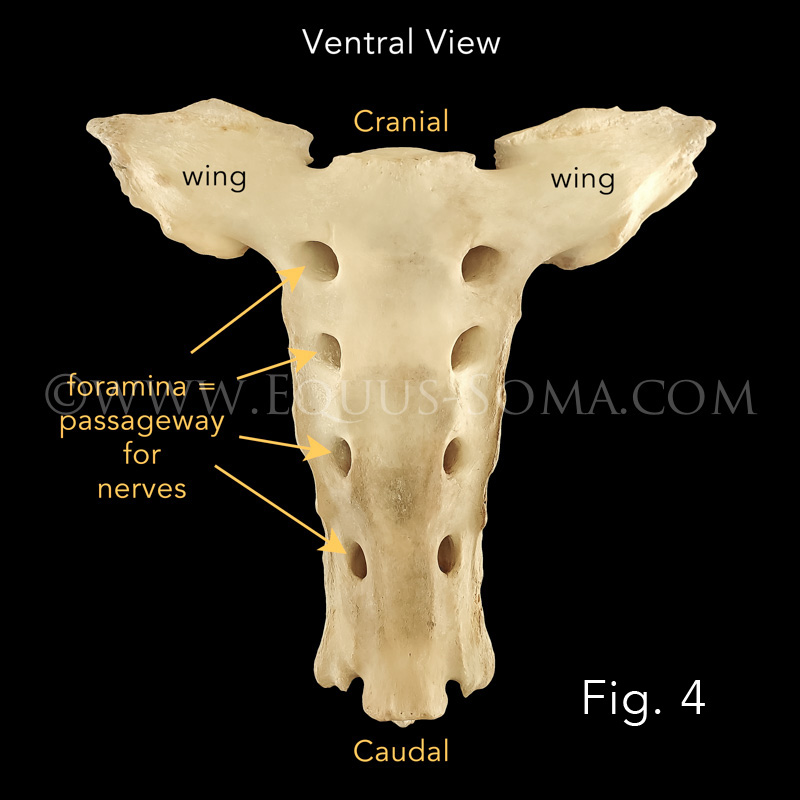
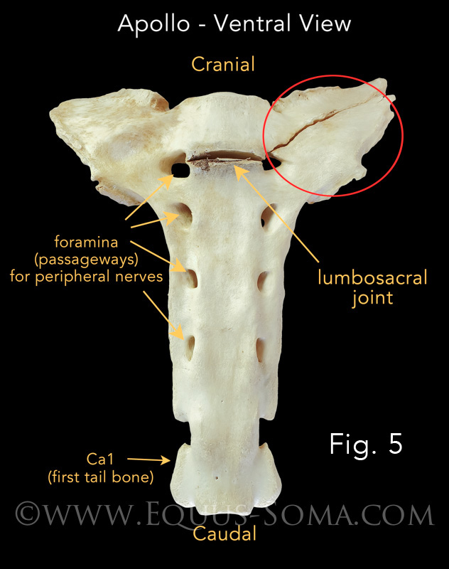
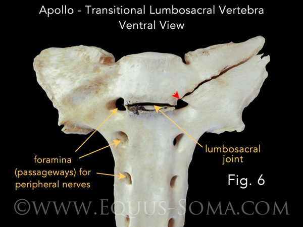
PHOTO CREDITS: The majority of images used on this website are property of Equus-Soma (Pamela Blades Eckelbarger). Images of me taken at Presentations are provided courtesy of Helen Peppe and other attending participants (thank you!!). Images on the About page of myself competing with Irish are courtesy of Flatlandsfoto. Images of skeletons in the banners are from Muybridge 1881.
November through July
1165 Shaws Fork Rd.
Aiken, SC 29805Equus-Soma
Equine Osteology & Anatomy Learning Center
Pamela Blades Eckelbarger M.S. Zoology
eqsoma71@gmail.com
(207) 542-61322024 ©ALL RIGHTS RESERVED
August through October
190 Horscents Ln.
Waldoboro, ME 04572