• Aiken, SC

    info@equus-soma.com

     
  • Equus-Soma

    Equine Osteology & Anatomy Learning Center

     
  • Waldoboro, ME

    207-542-6132

  • Apollo's Story - What Lies Beneath - Part Three

     **Please be sure to read Part One and Part Two of Apollo's Story to fully understand and appreciate what is described here in Part Three.**

  • Thoracolumbar Region


  •         
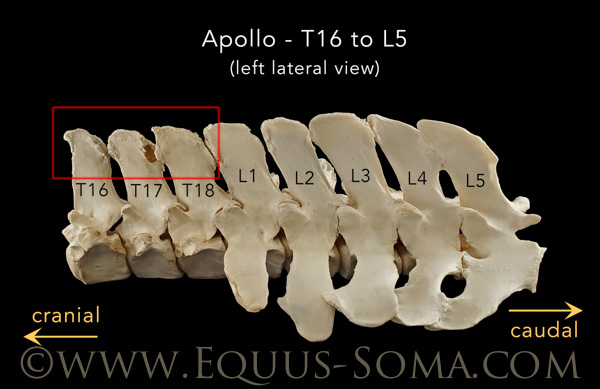
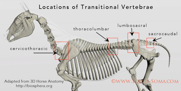
             Moving forward from the first lumbar vertebra (L1) in Apollo's lumbosacral region (discussed in Part Two) we will now look at the 18 vertebrae in Apollo's thoracic and thoracolumbar regions.

  •          The three photos below show left lateral views of T1 to T11, T11 to T18 (saddle region) and T16 to L5 (thoracolumbar region).   Unlike the lumbar vertebrae with their characteristic, wing-like transverse processes, T1 through T18 each carry a pair of ribs (not shown in the photos but illustrated in the diagram above).

    **CLICK ON ANY IMAGE TO ENLARGE**

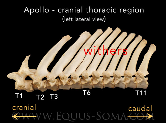
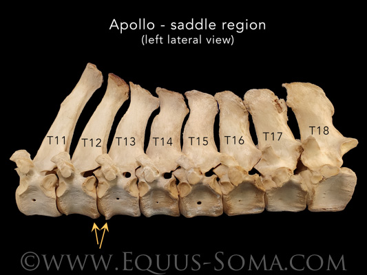
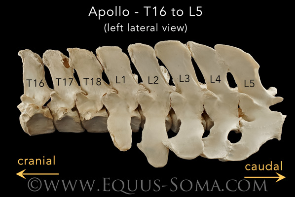
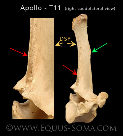
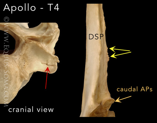
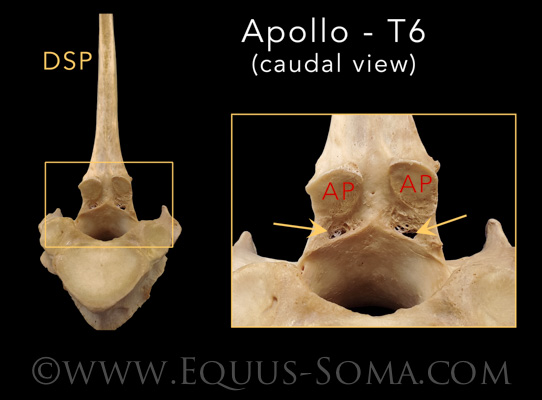
  •         Note the variations in size, shape and orientation of the dorsal spinous processes (DSP) from one region to the next.         
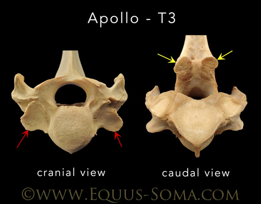
            The size, shape and orientation of the articular processes (APs) also vary between regions and have been reported to be biomechanically significant (see Apollo refs. #16 & 17).
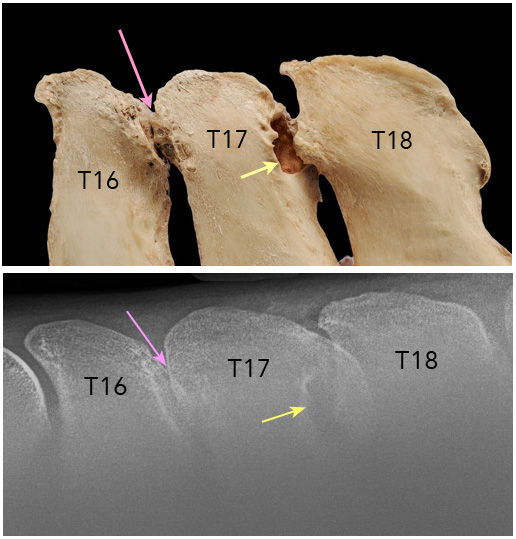
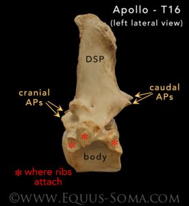
            The photo to the left is of Apollo's 16th thoracic vertebra (T16) illustrating the general morphology and sites where the ribs attach (costal fovea or costal facets) red astrisks.

             All of Apollo's thoracic vertebrae were closely examined for signs of mild, moderate or severe osteopathic lesions (as per Apollo refs 1, 3, 17 & 18).  In general, most of his cranial and mid-thoracic vertebrae were relatively "normal" in that they presented with low grade lesions that, though not insignificant, are routinely found in the bone collections here at the Learning Center and have been reported in post-mortem skeletal studies (see previous refs).

  •         The most pronounced osseous lesions in Apollo throughout all of his axial skeleton were present in the thoracolumbar region, specifically T16-L1 (red box in the photo below) and of course the lumbosacral region previously reviewed in Parts One and Two.

  •         The photo below compares an enlarged view of the T16, 17 and 18 DSPs (designated in the red box above) with a radiograph taken of the same DSPs.  The pink and yellow arrows point to the lesions resulting from impingement.  T17 has a deep lytic "hole" that probably formed from repetitive contact with the bony proliferation on the adjacent surface of T18.   According to Clayton & Stubbs (2016) these are "grade 3 lesions with lysis, proliferation and pseudoarticulation formation."

  •          It is very important to understand that even though we are talking about the bones in this study, the spine is surrounded and supported by musculature, ligaments, tendons, nerves and blood vessels all of which are interconnected through the fascial network.
             Above and between the DSPs are the supraspinous (SSL) and interspinous (ISL) ligaments which are highly innervated (see Apollo ref. #21).  Using techniques of histology, electron microscopy, and immunohistochemistry these authors provided a detailed description of the morphology of the SSL & ISL as well as associated connective tissue, musculature and fascia.  They stated in the discussion that "The dense sensory innervation within the SSL and ISL could explain the severe pain experienced by some horses with impinging dorsal spinous processes."

  •         The photos below illustrate other lesions (mostly mild) noted on Apollo's thoracic vertebrae cranial to T16.  These mainly include enthesophytes and periarticular osseous proliferation around some of the APs and costal fovea resulting in left-right asymmetry of these joints . 
            Enthesophytes  are osseous proliferations at sites where ligaments or tendons attach to the bone and are a result of inflammation of the connective tissue.  With respect to Apollo, only mild enthesophytes were seen on the DSPs of T11 and T4.

  •         In the photo below of T11, the image on the left is an enlarged view of the image on the right of the caudal edge at the base of the DSP.  The red arrows point to where the interspinous ligament (ISL) attached and the blade-like enthesophyte that formed there (as per Clayton & Stubbs, 2016).
            The green arrow indicates another mild enthesophyte on the cranial surface of the T11 DSP.

  •         In the photo below of T4, the image on the left is a close up of a costal fovea where part of the 4th rib attaches. Note the rough edge (osseous proliferation) and what appears to be a cleft or possible stress fracture (red arrow).
            The double yellow arrows indicate pointed, sharp enthesophytes along the caudal edge of the DSP.  The single yellow arrow indicates asymmetry of the caudal APs.

  •          Another example of asymmetry with extra osseous proliferation is shown in the photo below of Apollo's third thoracic vertebra (T3).  Note the different sizes of the costal fovea where part of the 3rd rib would attach (red arrow in cranial view).
             On the right image (caudal view), the yellow arrows point to the caudal articular process (AP) which are also asymmetrical in size and shape.

  •         The below photo shows the caudal view of the APs on Apollo's T6 vertebra.  The area in the yellow square is enlarged on the right illustrating both asymmetry in the size and shape of the APs, but also what appears to be lytic lesions between each AP and the dorsal arch (yellow arrows).
            Similar lesions were described by Clayton & Stubbs (2016) and assigned a grade 3 or severe rating.  Less severe lytic lesions are also present in the same area on Apollo's T7 and T8.