• Aiken, SC

    info@equus-soma.com

     
  • Equus-Soma

    Equine Osteology & Anatomy Learning Center

     
  • Waldoboro, ME

    207-542-6132

  • Apollo's Story - What Lies Beneath - Part Four

    ***Please be sure to read Part OnePart Two and Part Three of Apollo's Story prior to Part Four.***

  • Cervicothoracic Region

  •         Examination of Apollo's cervical (neck) vertebrae did not reveal much in the way of "major" pathology.  We did, however, find a number of asymmetries and osseous lesions that may be categorized as mild, moderate and a few possibly severe according to the detailed definitions provided by Haussler et al. (2019).

  •         We will begin with a general overview of "typical" cervical morphology, then illustrate the anomalies found in Apollo's cervicothoracic region.

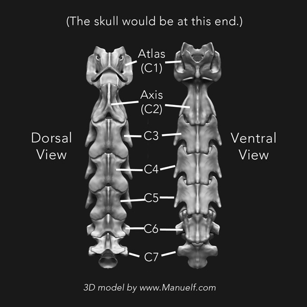
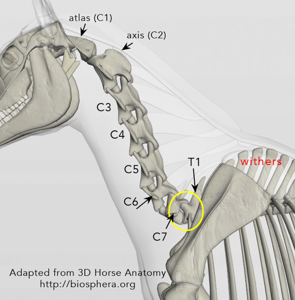
  •         Seven vertebrae make up the neck portion of the horse's spine and are designated "C1 thru C7".  C1, also known as the atlas, connects to the back of the skull at the atlantooccipital joint (poll).
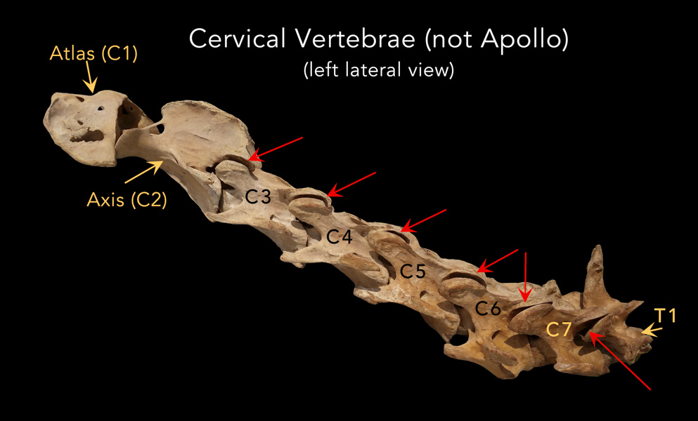
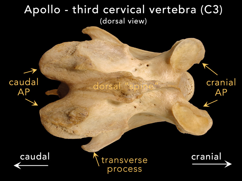
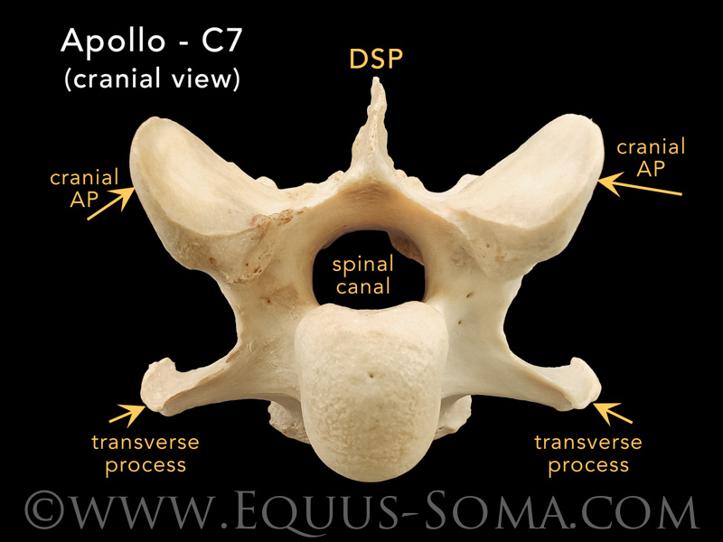
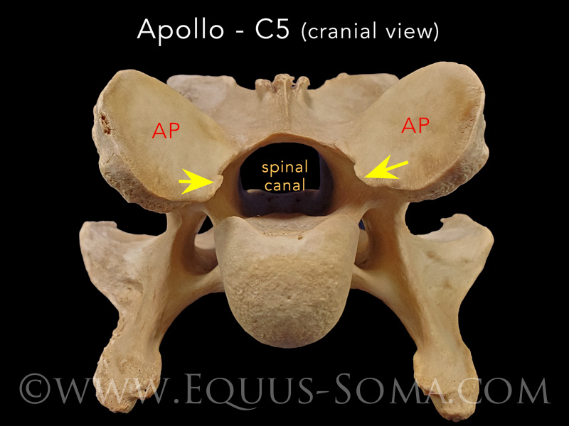
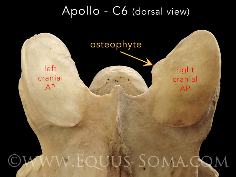
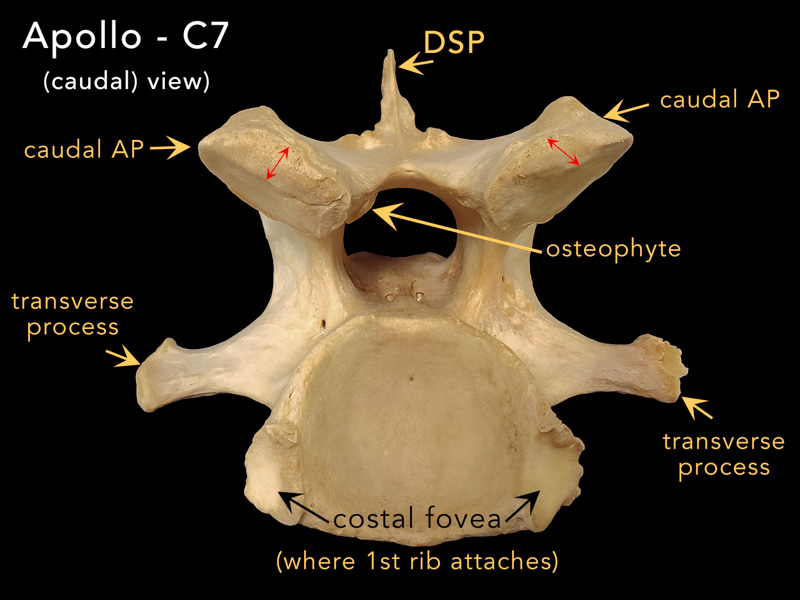
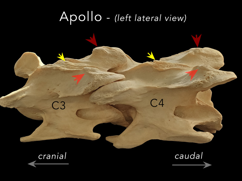
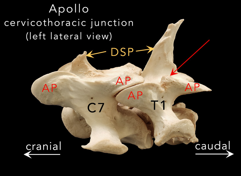
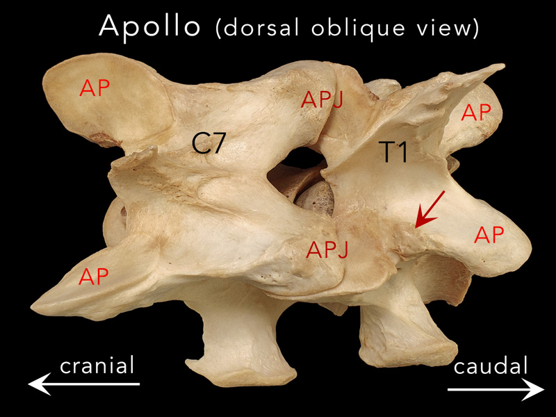
            The atlas (C1) and its immediate caudal neighbor the axis (C2) each have unique morphology compared to the remaining cervical vertebra.  C3, C4 and C5 are similar to each other.  C6 has a pair of distinct long "ridges" or "tubercles" on the ventral surface.  You can read more about C6 & C7 morphology (and their malformations) here.

            In the standing position there is an S-shaped curvature with a dorsal convexity at the poll and a ventral convexity at the C7-T1 junction.
            Both junctions are regions of large ranges of motion in flexion and extension. (see Apollo refs #23 & 24)

  •         The photo at right shows a lateral view of the 7 cervical (plus the first thoracic (T1)) vertebrae from another horse in the Learning Center collection. The skull would be connected to the atlas on the left of the image and the rest of the thoracic vertebrae would extend from T1 off to the right.
            The red arrows indicate the articular process joints (APJ) between each vertebra.
    ***CLICK ON PHOTO TO ENLARGE***

  •          Each cervical vertebra is comprised of a central body and dorsal arch through which passes the spinal canal.  Paired transverse processes run along the sides.  A pair of cranial articular processes (APs) and a pair of caudal APs extend from the respective ends on the dorsal surface. The dorsal spine is reduced to being more ridge-like except on C7 where a spine (DSP) of varying lengths may be present.

  • Osseous Changes to Apollo's Cervical Vertebrae

  •           Osseous anomalies found on Apollo's cervical vertebrae included osteophytes, AP "thickening", enlarged vascular channels, AP asymmetry, enthesophytes and an "extension impingement".   Most of the lesions were graded as "mild" but a few appeared to be "moderate to severe" as defined by Haussler et al. (2019).

  • Osteophytes

             Outward bony projections from the articular surface and margins of synovial joints are called “osteophytes”.  They are abnormal  bony outgrowths within the joint complex.  Osteophytes were found on some (not all) of the APs in Apollo's cervical vertebrae the majority of which were graded as "mild" to "moderate".
             Below: Mild osteophytes were present on the medial edge of the cranial APs of Apollo's C5 (yellow arrows) and C6. They were in close proximity to the spinal canal, but probably not large enough to invade the dural space.

  •         Below:  Moderately sized osteophytes were also present on the medial edges of both the left and right cranial APs of C3. The green arrow points to the medial edge of the right AP.  It is possible that these osteophytes may have reduced the size of the intervertebral foramina (yellow circle) through which cervical nerves pass as they exit the spinal cord.

  •         Below:  A moderately sized osteophyte was noted along the medial edge of the left caudal AP of C7. This bony projection does appear to invade the spinal canal just enough to have possibly caused issues with the spinal cord, especially during extreme ranges of extension.   Note also the "thickened" caudal edges of C7's caudal APs (double-headed red arrows)

  • Enthesophytes

              Enthesophyte formations are also present on some of Apollo's cervical vertebrae.  Unlike osteophytes which are bony projections that form at joint margins, enthesophytes (or bone spurs) form at entheses, the sites where tendons, ligaments, fascia, and joint capsules attach to bone.

             In other mammals, osteophytes are associated with aging and osteoarthritis, whereas, enthesophytes are more commonly seen at sites of repetitive muscle motion.

             The pathophysiology of enthesophyte formation continues to be debated in the literature.  Regrettably, heading down that rabbit hole is beyond the scope of this review of Apollo's bones, however, a noteworthy theory that may be applicable to sport horses suggests that enthesophytes develop in response to mechanical loading and may be a result of "overuse injuries." (see Apollo ref# 26)

  •         The most prominent enthesophyte formations in Apollo cervical region are at a few of the insertion sites of the multifidus cervicis muscle, specifically on the dorsal surfaces of the caudal APs of C3 and C4  (also described by Haussler et al. (2019).
            The multifidus cervicis is a rather complex, cybernetic muscle that runs along the dorsal surface from C2 to the first thoracic vertebra, T1.  It consists of 3 bundles arranged in 3 layers. Fibers from each paired bundle attach to a spinous process (yellow arrowheads below right) then split laterally, attach to the dorsal surface of the next vertebra and blend into the joint capsule of a caudal AP after crossing 1-4 intervertebral joints (long red arrows below left). (see Apollo ref #25)

  •          Above: The raised areas on the caudal APs of C3 and C4 to which the red arrowheads point, are enthesophytes that formed at the attachment sites of the multifidus cervicis muscle. 

  • Extension Impingement
  •          The cervicothoracic junction, otherwise known as the "thoracic sling" or "thoracic inlet" is at the base of the horse's neck (circled in yellow in the diagram to the right).
             This is the joint between the last cervical vertebra (C7) and the first vertebra of the rib-carrying thoracic region (T1) and is a key transitional region where the more freely moving cervical vertebrae meet the more fixed cranial thoracic vertebrae.
             Below:  Two slightly different views of Apollo's C7 and T1 vertebrae joined together to form the cervicothoracic junction.  Note the juxtaposition of the APs of both vertebrae to form the AP joint (APJ).
             The red arrows point to a knob-like osteophyte present only on the left side of T1's cranial AP.  The osteophyte is caudal to the articulating surface.
             This is referred to as an "extension impingement" that may have formed due to stresses produced by continuous "end-of-range intervertebral extension" (Haussler et al., 2019).

  •          Is it possible that in Apollo, the sliding of the left caudal AP of C7 was subjected to an abnormal range of motion in the caudal direction (i.e., extension) influenced by the asymmetry of the caudal APs, which resulted in their "sliding" past the "normal" area of the articular surface (dotted line in the photo above)?
             Could this have resulted in the proliferation of extra bone i.e., the osteophyte to act as a barrier to prevent further extension (?)

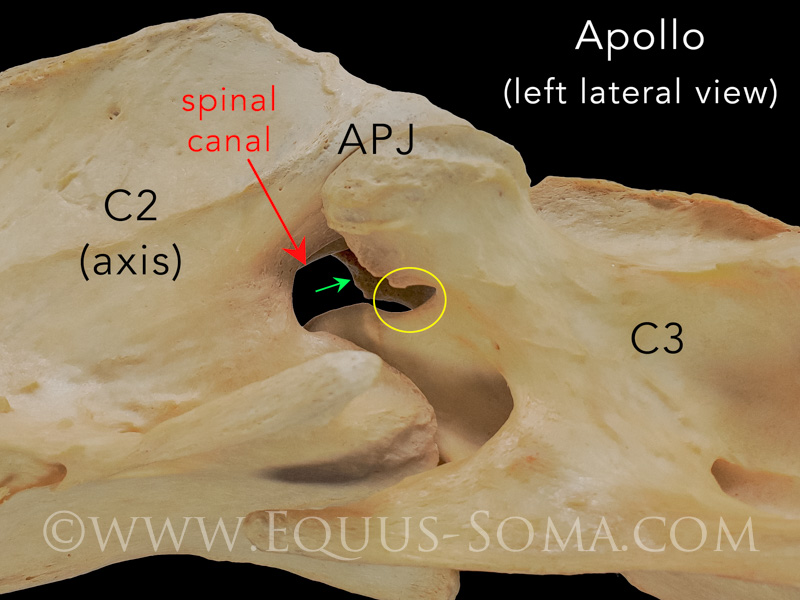
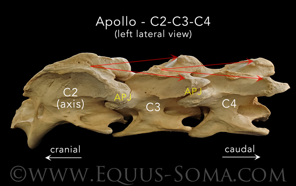
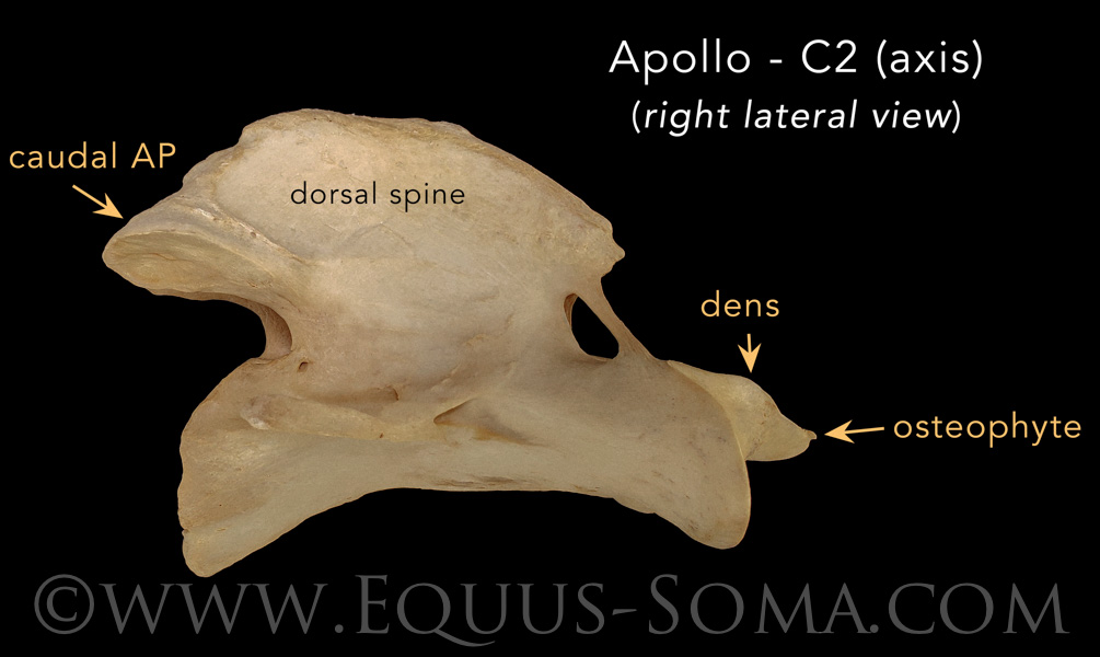
  •         With respect to Apollo's second cervical vertebra (C2, a.k.a. "axis") the only obvious bony lesion is a small osteophyte on the right lateral edge of the dens, which we consider to be of "mild" grade.

            On the other hand, Apollo's first cervical vertebra (C1, a.k.a. "atlas") presented with some very interesting lesions that we will elaborate on in Part Five where we describe his skull and the atlanto-occipital junction.