
Equus-Soma
Equine Osteology & Anatomy Learning Center
RESEARCH .... DISCOVERY .... EDUCATION
Aiken, SC
info@equus-soma.com
Waldoboro, ME
207-542-6132
ECVM Morphological Variations (Variants)
Before reading about the morphologies of the ECVM bones, please review the characteristics of the 'normal" C6 and C7 here.
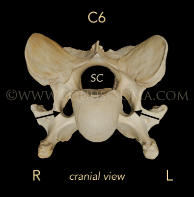
ECVM may involve C6 with or without C7, the 1st and 2nd sternal ribs, as well as the soft tissues that attach to these locations. In addition, the replication of one or a pair of foramen transversarium on C7 may occur (May-Davis et al., 2024 in review). The foramen transversarium (aka transverse or arterial foramen) are normally present on either side of the spinal canal (SC) in C1 to C6 (black arrows in photo to the right) but they are NOT present on normal C7. They provide channels through which the vertebral artery passes from the heart to the brain (Sisson & Grossman, 1975).
In some horses afflicted with the cervical malformation, one or both of the 1st sternal ribs may be missing or underdeveloped. Sharon also published a paper on the 1st sternal rib malformation (2017). More recently, and of growing concern, is the discovery that one or both of the 1st sternal ribs may be malformed/rudimentary when C6 has normal morphology (Ros et al, 2023; personal findings via dissections or skeletal remains).Changes in normal morphology of the vertebrae may affect the attaching musculature which may lead to disruption of normal balance and biomechanical function of the horse's neck, thoracic sling and ultimately through compensation, the whole body. (See Rombach et al, 2014; and May-Davis & Walker, 2015.)
**You are encouraged to download the pertinent publications as they contain more detailed information about all of these aspects of ECVM.**
Normal C6 - Cranial View
The congenital malformations to the C6 vertebrae are thought to involve faulty embryological development of portions of either the right, the left, or both ventral processes. Examining the morphology of C6 bones, we found that the quantity of missing bone can differ between the left and right ventral processes, even within a single horse. This led to the development of a grading system that identifies the extent of the absence (May-Davis et al., 2023).
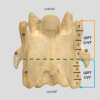
For grading purposes, we visualized dividing the cranial ventral tubercle (CrVT) and the caudal ventral tubercle (CVT) of each ventral process into relatively equal quarterly increments (photo to left, ventral view). As the grade number increases from 1 to 4, so does the amount of absent bone from the tubercle in the caudal to cranial direction. For example, when an absent caudal ventral tubercle (aCVT) displays a Grade 1, it indicates that 1/4 of the CVT is absent at the caudal-most region of that tubercle. At the opposite extreme, when an aCVT is assigned Grade 4, the entire CVT is missing.Examples of these C6 variants are illustrated by the 3D models below.
How to View the 3D Models
Click/tap on the 3D model of interest below. To see the model in full screen, click/tap on the double arrows in the lower right corner.
To move and rotate the model, grab it with the mouse or fingers and drag around. If you use a mouse with a scroll wheel, this will zoom in and out. Escape to exit full screen. On a tablet, two fingers to zoom, swipe down to exit full screen.
Numbers on the model, indicate annotations that explain the underlying structure. Click/tap on the number and a box with the description will open.PLEASE NOTE: Our paper described not only the grades of absent CVTs (aCVT), but in some horses we found that the missing tubercle extended further cranially to involve portions of the CrVT as well. Therefore the paper also includes grading for the absent CrVT (aCrVT) when present.
Since the bony changes on the aCrVT are more subtle and not easy to label in the 3D models, they will not be identified here. Please refer to the publication for full details.
**These 3D models are for on-line viewing only. The files cannot be downloaded.**
ECVM Variants - Unilateral C6
A "C6 unilateral" variant means that a portion of the left or the right caudal ventral tubercle did not develop. Return here to compare the unilateral variants with the normal C6 morphology.
Left Unilateral
- Right unilateral
The ventral morphology of the C6 to the right shows that the left CVT is slightly truncated. May-Davis et al., (2023) would assign a Grade 2 to this left CVT.
It is possible however, that the truncation of the left CVT may be due to separation of the ossification center (specific to C6) before it fully fused with the ventral tubercle.
In either scenario, would this shortened tubercle influence the portion of longus colli that normally anchors there?- Left unilateral - Grade 2 CVT?
ECVM Variants - Bilateral C6
A "C6 bilateral" variant means that a portion of BOTH the left and the right CVTs did not develop. The amount of bone that did not develop may differ between left and right CVTs, even within the same horse. As can be seen in the 3D model below (left).
The montage below (right) illustrates the bilateral C6 variants in our collection. This link will take you to the 3D models where you can select each to examine. Make note of the subtle differences in morphology.
Bilateral with asymmetrical CVTs
**Something Important to think about while viewing the ECVM C6 BILATERAL vertebrae**
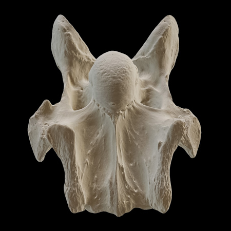
Examine the 3D models below of two different horses (Warrior and Swt). The C6 of both have left and right Grade 4 aCVTs, i.e., bilateral, completely missing caudal ventral tubercles. Compare these to the corresponding morphology of the C5 for each horse.
In the Grade 4 bilateral variation, C6 has acquired the morphological identity of C5.
1) Consider the FUNCTIONAL ramifications in the lower neck region.
2) How does this appear on radiographs? Can you or your veterinarian correctly identify C5 and C6 when the latter has the complete bilateral variant and they look nearly identical?This PDF was created to help owners and veterinarians better understanding how ECVM variants might appear in radiographs.
Warrior - Bilateral C6
Warrior - Normal C5
Swt - Bilateral C6
Swt - Normal C5
Coming Up Next
C7 variants in 3D
C7 - ECVM Variants
Rudimentary ribs
1st & 2nd Rib Malformations
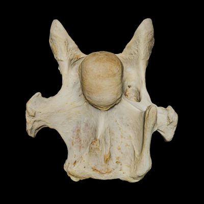
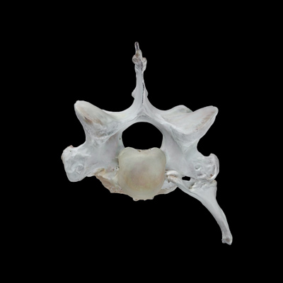
PHOTO CREDITS: The majority of images used on this website are property of Equus-Soma (Pamela Blades Eckelbarger). Images of me taken at Presentations are provided courtesy of Helen Peppe and other attending participants (thank you!!). Images on the About page of myself competing with Irish are courtesy of Flatlandsfoto. Images of skeletons in the banners are from Muybridge 1881.
November to July
1165 Shaws Fork Rd.
Aiken, SC 29805Equus-Soma
Equine Osteology & Anatomy Learning Center
Pamela Blades Eckelbarger M.S. Zoology
eqsoma71@gmail.com
(207) 542-61322025 ©ALL RIGHTS RESERVED
July through October
190 Horscents Ln.
Waldoboro, ME 04572