Aiken, SC
info@equus-soma.com
Equus-Soma
Equine Osteology & Anatomy Learning Center
Waldoboro, ME
207-542-6132
Superficial Dorsal and Superficial Ventral Lines
The Superficial Ventral Lines (SVL) and the Superficial Dorsal Lines (SDL) both connect at the head where the fibers of the Temporal muscle merge with the Masseter muscle, connecting the Temporomandibular joint (TMJ) through the length of the body.
The SVL and SDL also connect at the coffin bone (P3) of the hind limb where the extensor and flexor tendons attach (respectively), as well as through the branches of the Suspensory ligament as it merges with the extensor tendons.
Working together, the Superficial Ventral and Dorsal Lines coordinate flexion of the spine and hips and extension of the hind legs. If the SVL is hypertonic and the spine hyper-flexed, the hind limbs will be standing under.
The SDL influences the following musculoskeletal parts:
- Suspensory ligament of hind limbs (area of origin)
- Flexor tendons
- Gastrocnemius m.
- Biceps femoris m.
- Semitendinosus m.
- Sacrotuberous ligament
- Erector spinae muscle group (Spinalis m., Longissimus dorsi m., Iliocostalis m.)
- Longissimus cervicus m.
- Longissimus capitis m.
- Semispinalis capitis m.
- Ending at the crest of the Occiput & Temporal myofascia
Superficial Dorsal Line (SDL)
")
The SVL influences the following musculoskeletal parts:
- Extensor tendons, hind limb attachment @ P3 (origin) - Long & Lateral digital extensor mm.
- Peroneus tertius m.
- Patella & assoc. ligaments
- Quadricep femoris muscle group (Vastus lateralis m., V. medius m., V. intermedius m., Rectus femoris m.)
- Coxa-femoral joint
- Rectus abdominalis m.
- Rectus thoracis m.
- Sternum / manubrium of Sternum
- Sternomandibularis m.
- Mandible & ends in fascia of the Masseter m.
Superficial Ventral Line (SVL)
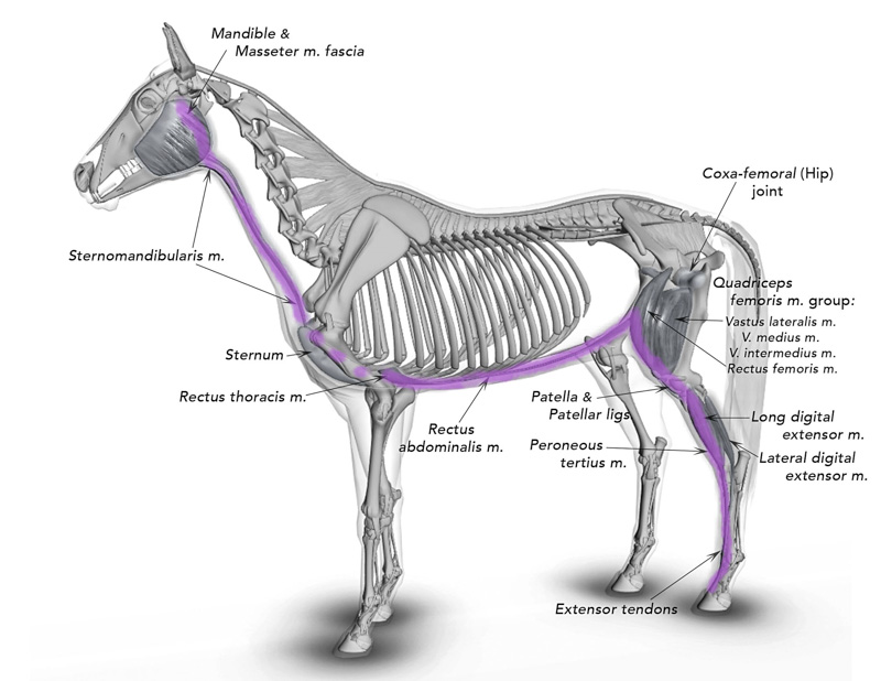
PHOTO CREDITS: The majority of images used on this website are property of Equus-Soma (Pamela Blades Eckelbarger). Images of me taken at Presentations are provided courtesy of Helen Peppe and other attending participants (thank you!!). Images on the About page of myself competing with Irish are courtesy of Flatlandsfoto. Images of skeletons in the banners are from Muybridge 1881.
November through July
1165 Shaws Fork Rd.
Aiken, SC 29805Equus-Soma
Equine Osteology & Anatomy Learning Center
Pamela Blades Eckelbarger M.S. Zoology
eqsoma71@gmail.com
(207) 542-61322024 ©ALL RIGHTS RESERVED
August through October
190 Horscents Ln.
Waldoboro, ME 04572