• Aiken, SC

    info@equus-soma.com

     
  • Equus-Soma

    Equine Osteology & Anatomy Learning Center

     
  • Waldoboro, ME

    207-542-6132

  • Bone Growth and Skeletal Maturation

             The complex and multi-faceted details of bone development and growth (ossification) are beyond the scope of this website but can be found in any book about embryology or bone biology.   The intention here is to combine a simplified overview of the process with images of bones from young horses in our Collections in order to substantiate previous reports that the skeleton of the horse is not fully mature at the ages most owners/trainers & riders want to start them.

              Ossification involves 2 distinct processes depending on the type of bone being formed.  Flat bones such as found in the skull, lower jaw and parts of the pelvis are formed by intramembranous ossification.  The axial (neck, back & pelvis) and appendicular (limbs) skeletons develop by means of endochondral ossification which involves cartilage.

              With a few exceptions (ex. the femur), the long bones (appendages) develop from 3 "centers of ossification".  The primary center develops the shaft of the bone (diaphysis) and 2 secondary centers of ossification are each associated with the ends of the bone (epiphysis).      

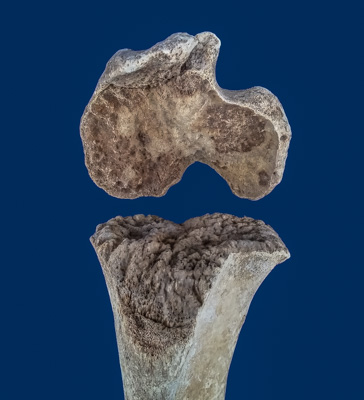
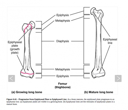
  •         The long bones lengthen as a result of gradual ossification within a layer of actively growing, embryonic cartilage (growth or epiphyseal cartilage) that occupies the space between the diaphysis and epiphyses.

            This thin layer of proliferating cartilge is called the epiphyseal plate or GROWTH PLATE.  As long as this cartilage continues to grow, new bone will be formed at the ends, thus increasing the length of the bone.

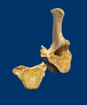
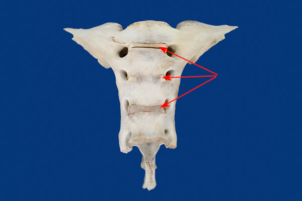
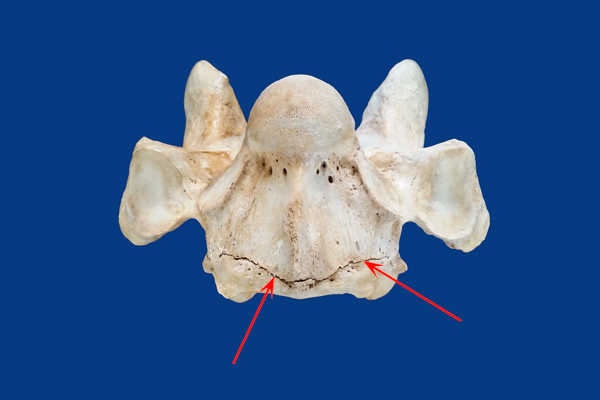
            Vertebral  bodies i.e., the bones that make up the the neck, back and sacrum, develop by endochondral ossification within cartilage that surrounds the notochord and forms the sides of the neural canal.  Each vertebra has 5 "centers of ossification";  3 are primary centers (form the body and arches that border the spinal canal) and 2 are secondary centers of ossification. These are the thin physeal plates at the cranial (towards the head) and caudal (towards the tail) ends of the main vertebral body (centrum).

  •        FUSION = MATURATION
              When the epiphyseal cartilage stops growing, it undergoes a final ossification and the result is the "fusion" of the epiphyses to the shaft of the long bone, and the physeal endplates to the centrum of the vertebra.

    Therefore, horse's bones are not structurally mature until after this final fusion is complete.

  •           Herein lies the confusion.  At the mention of "fusion of the growth plates" most people think this refers to the horse's knees ... "wait until the kness close".  What is not fully understood is that all of the bones in the skeleton have multiple epiphyseal (cartilagious) growth plates and that all of these must go through the final ossification step of fusion before the whole skeleton becomes structurally mature. 

             Furthermore....  the rate at which this fusion process occurs varies between bones and within the skeleton as a whole.  Look at the Fusion Timeline in the sidebar and notice that the growth plates from the knees down fuse before the other long bones.  PLUS, the LAST bones to undergo final fusion of the growth plates are the vertebrae of the neck and back. 

             Even more importantly, according to Dr. Deb Bennett (see Sidebar for PDF) the very last bones of the skeleton to close are the 7th cervical and 1st thoracic (C7-T1 junction), i.e., at the BASE OF THE NECK.

  • Click on the boxes below to view the IMMATURE bones of horses 5 years old and under...